How to Turn an Eye Exam Into a Better Everyday Vision Plan
How to Turn an Eye Exam Into a Better Everyday Vision Plan Effective routine eye care becomes more useful when patients connect clinical testing with…

Effective routine eye care becomes more useful when patients connect clinical testing with the actual places, distances, and tasks where their vision needs to perform.
An eye exam can measure clarity, update a prescription, and evaluate ocular health. Yet the quality of the final plan also depends on information that no instrument can collect by itself. How far away is the computer monitor? Does blur begin after twenty minutes or six hours? Is night driving difficult because of glare, poor distance clarity, or a scratched lens?
These practical details help turn measurements into decisions. Preparing them in advance can make the conversation more precise without requiring patients to understand clinical terminology.
Most people use their eyes across a wider range of distances than “near” and “far.” A typical day may include a phone, laptop, desktop monitor, conference-room screen, dashboard, grocery shelf, television, and nighttime road signs.
Before an exam, identify the tasks that matter most:
Estimate the usual working distance for the highest-priority tasks. A laptop positioned close to the body creates a different visual demand from a large monitor placed farther away. These differences can influence discussions about lens design and working posture.
“My vision is blurry” is an important starting point, but a pattern provides more useful information. Note which eye seems affected, when the problem occurs, how long it lasts, and what improves it.
A simple symptom log can record:
This level of detail helps distinguish a focusing or prescription complaint from a surface issue, binocular-vision concern, medication effect, or symptom requiring a different level of urgency.
Health history matters because the eyes do not operate separately from the rest of the body. Bring an accurate medication and supplement list, including recent changes. Report diabetes, high blood pressure, autoimmune conditions, allergies, migraines, previous eye injuries, and earlier procedures.
Family history should be specific whenever possible. “Poor eyesight” might mean ordinary nearsightedness, while a documented history of glaucoma, retinal detachment, or macular degeneration may influence risk assessment.
Also bring glasses used for different activities, even if they are old or rarely worn. The clinician can learn from what works, what fails, and how previous prescriptions have changed.
Refraction determines which lens powers improve focus. The National Eye Institute explains that myopia, hyperopia, astigmatism, and presbyopia occur when the eye does not focus light correctly on the retina.
The familiar “which is clearer, one or two?” comparison is subjective by design. It incorporates the patient’s visual response into the measurement. Taking time to answer consistently is more useful than trying to guess which response seems correct.
Refraction does not, by itself, describe the health of the retina, optic nerve, cornea, lens, eyelids, or tear film. A comprehensive evaluation combines prescription testing with other procedures selected according to the patient’s needs.
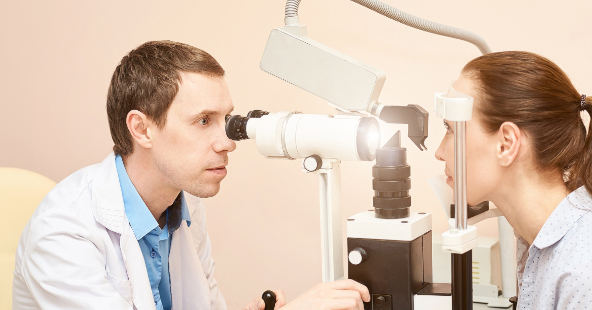
Dilation temporarily widens the pupils so the doctor can view more of the eye’s internal structures. The National Eye Institute’s dilated exam guide explains that dilation supports evaluation for conditions including diabetic retinopathy, glaucoma, and age-related macular degeneration.
Close vision may be temporarily blurred, and light sensitivity can last for several hours. Bring sunglasses and ask the office what to expect if driving or detailed work is planned afterward.
Retinal images can document visible structures, but an image should not be treated as a self-interpreting photograph. Image quality, field of view, symptoms, history, and the rest of the clinical examination influence what a finding means.
Long periods of computer use can be associated with blur, dryness, burning, headache, and neck or back discomfort. A CDC Stacks resource on computer vision syndrome recommends regular visual breaks, workstation adjustments, glare reduction, suitable screen brightness, and regular eye exams.
For a more productive discussion, record:
These observations help separate an optical correction issue from environmental dryness or workstation design. More than one factor may be present.
Routine care should address how the eyes are used and protected. Prescription glasses are not automatically safety eyewear.
The National Institute for Occupational Safety and Health recommends matching eye protection to the nature and extent of the hazard while considering coverage, comfort, peripheral vision, and other protective equipment.
Tell the eye doctor about exposure to dust, chemicals, flying particles, biological material, impact, or intense light. The same principle applies to home repairs, yard work, hobbies, and sports. Clear vision and adequate protection need to be planned together.
Contact lens wearers should bring their current lens details and describe actual habits rather than the routine they intended to follow. Wearing time, sleeping, showering, swimming, replacement timing, solutions, and case care all affect the ocular surface.
The U.S. Food and Drug Administration advises keeping contacts away from water, using fresh solution, following the recommended schedule, and contacting an eye-care professional when irritation or infection symptoms develop.
Bring backup glasses and remove contacts when pain, redness, unusual discharge, light sensitivity, or sudden blur occurs. Continuing to wear a lens through significant discomfort can make evaluation more difficult and may worsen a problem.
At the end of the visit, the findings should lead to an understandable plan. Ask:
Patients are entitled to their eyeglass prescription. The Federal Trade Commission’s Eyeglass Rule requires optometrists and ophthalmologists to provide a copy after a refractive examination without an additional fee. That prescription preserves consumer choice, while professional lens measurements and fitting remain important to the finished eyewear.
A scheduled exam is not the correct pathway for every symptom. Sudden vision loss, severe eye pain, chemical exposure, significant injury, or a rapid increase in flashes and floaters may require urgent or emergency evaluation.
The National Eye Institute identifies sudden new floaters, flashes of light, and a curtain-like shadow as possible retinal-detachment symptoms that require immediate care.
When contacting an office, state the symptom first, when it began, and whether one or both eyes are affected. This helps the clinical team determine the appropriate response.
Nova Optique & Eyecare in Arlington, Virginia, states that its annual comprehensive exam includes evaluation of the eyes, lashes, and lids, an eyeglass prescription, and retinal images. Its published information also identifies glaucoma, macular degeneration, and retinal holes or tears among the ocular issues assessed.
The practice notes that a comprehensive exam generally takes thirty minutes to one hour depending on the patient’s needs. This variable timeframe reflects an important reality: useful care is guided by the questions and findings present, not merely by completing the same sequence at the same speed for every person.
Patients do not need to diagnose themselves or arrive with technical vocabulary. They can improve the value of an exam by bringing accurate history, describing symptoms as patterns, identifying essential visual tasks, and asking what the results mean for daily life.
That preparation turns routine care into a practical planning process. The outcome is not just a set of measurements. It is a clearer understanding of current vision, relevant risks, appropriate correction, protective habits, and what should happen next.